Home
/ Lower Body Skeleton Diagram / Bones Of The Leg And Foot Interactive Anatomy Guide - Human skeleton, the internal skeleton that serves as a framework for the body.
Lower Body Skeleton Diagram / Bones Of The Leg And Foot Interactive Anatomy Guide - Human skeleton, the internal skeleton that serves as a framework for the body.
Lower Body Skeleton Diagram / Bones Of The Leg And Foot Interactive Anatomy Guide - Human skeleton, the internal skeleton that serves as a framework for the body.. Did you know… ☛ while the size of the human head right from birth won't change drastically, it is the torso and the lower limbs that grow in length. The skeleton protects vital organs. Abdominal anatomy pictures 12 photos of the abdominal anatomy pictures abdominal internal organs anatomy pictures, anatomy of abdominal cavity pictures, anatomy pictures abdominal organs, female abdominal anatomy pictures, human abdominal anatomy pictures, human anatomy, abdominal internal organs anatomy pictures, anatomy. Human skeleton, the internal skeleton that serves as a framework for the body. The heart and lungs are located within the thoracic cavity, and the vertebral column provides structure and protection for the spinal cord.
Related posts of anatomy of lower body abdominal anatomy pictures. The skeleton is divided into 2 main parts: These sections are cervical (neck), thoracic (upper and middle back), lumbar (lower back), and sacrum (tailbone). Abdominal anatomy pictures 12 photos of the abdominal anatomy pictures abdominal internal organs anatomy pictures, anatomy of abdominal cavity pictures, anatomy pictures abdominal organs, female abdominal anatomy pictures, human abdominal anatomy pictures, human anatomy, abdominal internal organs anatomy pictures, anatomy. Leg muscles anatomy muscular system anatomy human muscle anatomy anatomy bones human anatomy and physiology body anatomy leg muscles diagram muscle diagram lower leg muscles.
Skeletal System Lower Body Diagram Quizlet from o.quizlet.com Out of these, the cookies that are categorized as necessary are stored on your browser as they are essential for the working of basic functionalities of the website. The skeleton acts as a scaffold by providing support and protection for the soft tissues that make up the rest of the body. There also are bands of fibrous connective tissue—the ligaments and the tendons—in intimate relationship with the parts of the skeleton. Consists of the skull, ribs, vertebral column and sternum. Lower leg muscle diagram blank sketch coloring page. The sacroiliac (si) joints connect the sacrum at the base of the spine with the hip bone. Get product information, training, support, solutions and more. The bones of the pelvis and lower back work together to support the body's weight, anchor the abdominal and hip muscles, and protect the delicate vital organs of the vertebral and abdominopelvic cavities.
Altogether, the skeleton makes up about 20 percent of a person's body weight.
Posted on june 7, 2016 by admin. Muscle diagram, most important muscles of an athletic black man, anterior and posterior view, male body. The bones of the pelvis and lower back work together to support the body's weight, anchor the abdominal and hip muscles, and protect the delicate vital organs of the vertebral and abdominopelvic cavities. Your lower back (lumbar spine) is the anatomic region between your lowest rib and the upper part of the buttock. The human spine is composed of 4 sections of vertebrae. Learn how to communicate up to six times better by using visuals with smartdraw. Evenly distribute weights from your upper body into the lower extremities. This diagram depicts picture of female reproductive system diagram 1024×1204 with parts and labels. The brain is surrounded by bones that form part of the skull. Lower leg muscle diagram blank sketch coloring page. Related posts of anatomy of lower body abdominal anatomy pictures. Female anatomy includes the external genitals, or the vulva, and the internal reproductive organs. This curve, called lordosis, helps to:
The lumbar and sacrum region make up the bone of the lower back anatomy. It is made up of five larger vertebrae. Woman holding a blackboard with an illustration of the human digestive system drawn on it in chalk. Bones of the pelvis and lower back. The appendicular skeleton includes the bones of the shoulder girdle, the upper limbs, the pelvic girdle, and the lower limbs.
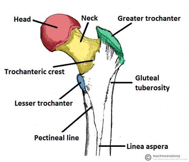
Bones Of The Lower Limb Teachmeanatomy from teachmeanatomy.info The appendicular skeleton includes the bones of the shoulder girdle, the upper limbs, the pelvic girdle, and the lower limbs. Abdominal anatomy pictures 12 photos of the abdominal anatomy pictures abdominal internal organs anatomy pictures, anatomy of abdominal cavity pictures, anatomy pictures abdominal organs, female abdominal anatomy pictures, human abdominal anatomy pictures, human anatomy, abdominal internal organs anatomy pictures, anatomy. The skeleton protects vital organs. The myology of the lower limb is also particularly well represented in this atlas of anatomy, with multiple anatomical charts and diagrams: There are 206 bones in the body which make up the skeleton. The pectoral muscles, the abdominal muscles, and the lateral muscle. The lumbar and sacrum region make up the bone of the lower back anatomy. Related posts of anatomy of lower body abdominal anatomy pictures.
Abdominal anatomy pictures 12 photos of the abdominal anatomy pictures abdominal internal organs anatomy pictures, anatomy of abdominal cavity pictures, anatomy pictures abdominal organs, female abdominal anatomy pictures, human abdominal anatomy pictures, human anatomy, abdominal internal organs anatomy pictures, anatomy.
Diagrams of human muscles lower arm muscles diagram human muscle. Did you know… ☛ while the size of the human head right from birth won't change drastically, it is the torso and the lower limbs that grow in length. The brain is surrounded by bones that form part of the skull. The bmi is defined as the body mass divided by the square of the body height. The skeleton acts as a scaffold by providing support and protection for the soft tissues that make up the rest of the body. The first diagram summarizes the different muscular compartments (fascial compartments) of the thigh and leg, and the different fascias (crural fascia, intermuscular septum, interosseous membrane, adductor canal, fascia lata) The main differences and dive into the anatomy and function of. Each bone is a complex living organ that is made up of many cells, protein fibers, and minerals. Lower leg muscle diagram blank sketch coloring page. There are 206 bones in the body which make up the skeleton. There also are bands of fibrous connective tissue—the ligaments and the tendons—in intimate relationship with the parts of the skeleton. The skeleton protects vital organs. Evenly distribute weights from your upper body into the lower extremities.
This article looks at female body parts and their functions, and it provides an interactive diagram. Together these bones form the vertical axis of the body (the axial skeleton). The skull, vertebral or spinal column (also known as the backbone or spine), breastbone (or sternum), and ribs are a major division of the skeleton; The skeleton is divided into 2 main parts: The human spine is composed of 4 sections of vertebrae.
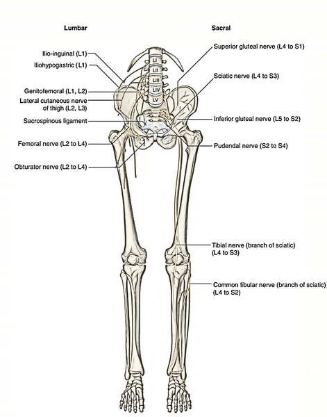
Easy Notes On Lower Limb Learn In Just 4 Minutes Earth S Lab from www.earthslab.com There are 5 different types of bones in the skeleton. Together these bones form the vertical axis of the body (the axial skeleton). Lower leg muscle diagram blank. Balance the weight of your head on top of your spine. This framework consists of many individual bones and cartilages. Human skeleton, the internal skeleton that serves as a framework for the body. Out of these, the cookies that are categorized as necessary are stored on your browser as they are essential for the working of basic functionalities of the website. It is made up of five larger vertebrae.
There are 5 different types of bones in the skeleton.
1 your spine in this region has a natural inward curve. Related posts of anatomy of lower body abdominal anatomy pictures. Lower leg muscle diagram blank. Learn vocabulary, terms, and more with flashcards, games, and other study tools. The torso of the human body also consists of the major muscles of our body; Posted on june 7, 2016 by admin. Let's take a look at the bones of the appendicular skeleton. These muscles help the body bend at the waist. The skeleton protects vital organs. This article looks at female body parts and their functions, and it provides an interactive diagram. Your lower back (lumbar spine) is the anatomic region between your lowest rib and the upper part of the buttock. This framework consists of many individual bones and cartilages. This diagram depicts picture of the female body 744×992 with parts and labels.
Consists of the skull, ribs, vertebral column and sternum lower body diagram. There also are bands of fibrous connective tissue—the ligaments and the tendons—in intimate relationship with the parts of the skeleton.
